The short answer
We are the leading provider nationally for artery and vascular scans. Our specialist vascular scientists complete a full screen of your major arteries, including the carotid, aorta, femoral, popliteal and pedal arteries, and an ankle brachial pressure index (ABPI) that compares the blood pressure in your arms and legs.
Vascular imaging is part of Ultimate (£1,299) and Ultimate+ (£1,799).
What is a vascular scan?
A completely non-invasive, ultrasound-based scan of your cardiovascular system. We use duplex imaging, which combines a picture of the artery with a measurement of the blood flowing through it, to see the health of your blood vessels and how well blood flows around your body. In Ultimate+, we add an ultrasound of the heart (an echocardiogram) to check how it works in detail.
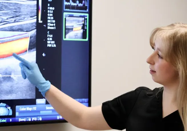
The arteries we scan
| Artery | Where | Why it matters |
|---|---|---|
| Carotid arteries | Neck | Plaque or narrowing here can lead to stroke |
| Aorta | Abdomen | We check for widening of the artery, an aneurysm |
| Femoral and popliteal arteries | Groin, thigh and behind the knee | Disease here can cause pain when walking and, at worst, gangrene or amputation. It is also a sign of higher heart attack and stroke risk |
| Pedal arteries | Feet | How well blood reaches the furthest points of your circulation |
| Ankle brachial pressure index (ABPI) | Arms and legs | Comparing blood pressure in your arms and legs shows whether circulation to the legs is reduced |
What we look for
We look at the health of your arteries and veins and how well blood flows through your body, to detect:
- blockages and blood clots;
- thickening of the artery wall;
- plaque;
- narrowing (stenosis);
- aneurysms;
- poor circulation.
Together these show your atherosclerotic burden: how much disease has built up in your arteries as a result of high blood pressure, cholesterol, diabetes or smoking. Risk factors tell us what could happen. Imaging shows whether it already is.
Why it’s different
- A moving picture, not a snapshot. Unlike CT or MRI, our scans show your blood flowing in real time, which helps us tell normal ageing apart from something that needs attention.
- No needles, no radiation, no downtime.
- Results on the day, explained by expert vascular scientists.
- See your own scan as we talk you through it.
How plaque forms
Plaque develops through a long, slow inflammatory process inside the artery wall, rather than cholesterol simply sticking to the surface. In simplified terms:
- EntryApoB-containing particles, such as LDL, pass through the artery’s inner lining into the wall. The more of them there are in your blood, the more chances this has to happen.
- RetentionSome particles become trapped in the wall and are chemically changed. The body recognises them as a problem.
- InflammationImmune cells move in, engulf the trapped fat and become foam cells. Together they form the earliest visible lesion, a fatty streak.
- PlaqueSmooth muscle cells build a fibrous cap over a fatty core. Calcium can build up over time.
- RuptureSome plaques stay stable for years. Others become inflamed and their cap thins. If it breaks, a clot can form quickly, and that clot, rather than gradual narrowing, is what can suddenly cause a heart attack or some types of stroke.
High ApoB and LDL, smoking, high blood pressure and diabetes all speed this process up. That is why we measure ApoB in your blood analysis: it shows how many particles are circulating that are capable of forming plaque. Cholesterol results explained.
What affects your ApoB
- Genetics: inherited variants, such as familial hypercholesterolaemia, can raise ApoB substantially.
- How well your liver clears particles: fewer working LDL receptors mean particles stay in your blood for longer.
- Insulin resistance: can make the liver release more particles, one reason ApoB is often raised with metabolic syndrome and type 2 diabetes.
- Diet and body composition: saturated fat can raise ApoB in some people, and replacing it with unsaturated fats generally lowers it. Excess energy intake and visceral fat can increase production.
- Other conditions: an underactive thyroid, kidney disease and some liver and metabolic disorders can raise ApoB.
- Medicines: statins, ezetimibe and other lipid-lowering treatments lower ApoB.
Inside the artery wall
An artery wall has three main layers around the lumen, the channel the blood flows through:
- Tunica intima (inner layer): the thin lining in direct contact with the blood. This is where ApoB particles cross in and plaque develops.
- Tunica media (middle layer): mainly smooth muscle and elastic tissue, which lets the artery constrict and widen and withstand the pressure from each heartbeat.
- Tunica adventitia (outer layer): connective tissue with nerves and, in larger arteries, tiny blood vessels that supply the wall itself.
Because plaque grows within the wall, it can thicken the wall for years before it narrows the lumen. Ultrasound can see that thickening and plaque early.
When to see a doctor instead
Call 999 if you notice signs of a stroke: the face drooping on one side, weakness in an arm, or slurred speech. Call even if the symptoms pass.
See your GP if you get pain in your calves, thighs or buttocks when walking that eases with rest, or a sore on your foot or leg that won’t heal.
Questions
Is a vascular scan painful?
No. It is a non-invasive ultrasound scan: you will feel cool gel and light pressure from the probe. There are no needles.
Does an artery scan use radiation?
No. Ultrasound uses sound waves, the same technology used in pregnancy scans.
Which Health Checks include artery imaging?
Ultimate (£1,299) and Ultimate+ (£1,799) include comprehensive vascular imaging and dynamic blood-flow analysis, alongside everything in the Health Check.
Why don’t you use calcium CT scoring?
A CT calcium score is a static image, taken at rest, of the heart’s blood vessels only. It shows how much disease is there, but not whether it is affecting how your heart works, and it says little about your risk of stroke, aneurysm or peripheral arterial disease. Our scans look at your arteries throughout your body in real time, without ionising radiation.